atlas of human anatomy pdf netter
Summary
Unlock the ultimate visual guide to human anatomy with Netter’s Atlas PDF. Your go-to resource for medical studies – download now from Sarasota CCW!

Netter’s Atlas of Human Anatomy is a renowned medical resource, offering detailed, clinically relevant illustrations. The 7th edition provides updated visuals, enhancing understanding of human anatomy.
Overview of the Atlas
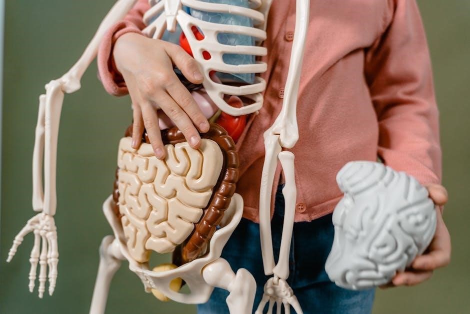
Netter’s Atlas of Human Anatomy is a leading medical resource, renowned for its detailed and clinically relevant illustrations. Created by Frank H. Netter, MD, it provides exquisite views of the human body, focusing on clarity and precision. The atlas is widely used in medical education and practice, offering a comprehensive understanding of anatomy from a clinical perspective. Available in multiple editions, including the 7th and 8th, it features updated content, enhanced visuals, and interactive tools like Netter’s Interactive Dissector. Its popularity stems from its ability to bridge basic science with practical applications, making it an essential tool for students and professionals alike. The PDF version is highly sought after for its convenience and accessibility, ensuring that Netter’s timeless work remains a cornerstone in anatomical education and reference.
Frank H. Netter and His Contributions
Frank H. Netter, MD, was a physician and illustrator whose detailed, clinically relevant artwork revolutionized medical education. His legacy includes the iconic Atlas of Human Anatomy.
Biography of Frank H. Netter
Frank H. Netter, MD, was a physician and illustrator whose detailed, clinically relevant artwork revolutionized medical education. His legacy includes the iconic Atlas of Human Anatomy, which remains a cornerstone in medical education. Netter’s unique ability to blend artistic talent with medical expertise created visual representations that are both accurate and accessible. His work continues to influence generations of healthcare professionals, solidifying his impact on the field of anatomy and beyond.
Editions of Netter’s Atlas of Human Anatomy
Netter’s Atlas of Human Anatomy has multiple editions, each offering updated illustrations and clinical correlations, with the 7th and 8th editions being particularly renowned.
7th Edition Features
The 7th edition of Netter’s Atlas of Human Anatomy features updated illustrations and clinical correlations, providing a comprehensive regional approach to human anatomy. It includes detailed views of the head, neck, and upper limb, along with enhanced depictions of neuroanatomy and systemic structures. The edition is designed for medical students and professionals, offering clear, concise descriptions. The PDF version is widely available, making it accessible for study and reference. Its focus on clinical relevance ensures practical applications in medical education and practice, solidifying its reputation as a cornerstone resource in anatomy.
8th Edition Updates
The 8th edition of Netter’s Atlas of Human Anatomy introduces enhanced illustrations and improved organization for better clarity. New clinical correlations and updated systemic content provide deeper insights. The regional approach remains central, with expanded coverage of complex anatomical structures. Digital access has been optimized, offering interactive tools like the Netter’s Interactive Dissector. These updates ensure the atlas remains a vital resource for medical education and practice, maintaining its legacy as a trusted reference for anatomy students and professionals worldwide.

Structure and Organization
Netter’s Atlas is organized regionally, covering areas like head, neck, and thorax. It also offers a systems-based approach, integrating anatomy with physiology for clarity and educational purposes.
Regional Approach
Netter’s Atlas employs a regional approach, systematically covering body regions like the head, neck, thorax, abdomen, and extremities. Each section provides detailed, clear illustrations, enhancing comprehension of complex anatomical relationships. This organization mimics how anatomical structures are encountered during dissection, making it ideal for hands-on learning. The regional focus also aids in correlating anatomy with clinical scenarios, a valuable tool for both medical students and professionals. Additionally, the atlas includes unlabeled figures for self-testing, reinforcing retention of anatomical knowledge effectively.
Systems-Based Approach
Netter’s Atlas also adopts a systems-based approach, organizing content by physiological systems such as the nervous, circulatory, and digestive systems. This method facilitates a deeper understanding of how anatomical structures function collectively. Detailed illustrations highlight relationships between organs and tissues within each system, aiding in the comprehension of complex physiological processes. The systems-based approach is particularly useful for students and professionals needing to grasp clinical correlations and diagnose conditions effectively. It complements the regional approach, offering a comprehensive learning experience tailored to various educational needs and preferences.

Clinical Applications
Netter’s Atlas is invaluable in clinical settings, providing clear visuals for identifying anatomical structures and understanding surgical procedures. Its precision aids healthcare professionals in diagnostics and treatment planning.
Use in Medical Education
Netter’s Atlas of Human Anatomy is a cornerstone in medical education, widely used by students and professionals alike. Its clear, detailed illustrations and clinical focus make it an essential resource for understanding human anatomy. The atlas is often integrated into curriculum, helping learners visualize complex structures and their relationships. The 7th edition, in particular, is praised for its updated visuals and concise descriptions, making it a go-to reference for both classroom and clinical training. Its ability to bridge anatomy with practical applications ensures its enduring popularity in medical education.

Downloading the PDF
Netter’s Atlas of Human Anatomy PDF is available for download in its latest editions, including the 7th and 8th editions, from legitimate sources online.
Legitimate Sources
To download the Netter’s Atlas of Human Anatomy PDF, ensure you use legitimate sources like the official Elsevier website or authorized retailers; Purchasing directly from the publisher guarantees access to the latest editions with high-quality illustrations. Additionally, many medical schools and libraries provide authenticated access to the digital version. Always avoid unauthorized websites to comply with copyright laws and ensure you receive the complete, updated content. Regular updates and new features in recent editions make legitimate sources the best choice for accurate anatomical knowledge.
Interactive Features
Netter’s Atlas offers interactive features like the Netter’s Interactive Dissector, providing 3D views and labeled structures for enhanced anatomical understanding and learning experiences.
Netter’s Interactive Dissector
Netter’s Interactive Dissector is a powerful digital tool offering 3D views of human anatomy. It allows users to explore structures layer by layer, enhancing learning. The tool features labeled illustrations, enabling precise identification of anatomical details. Ideal for medical students and professionals, it complements the atlas by providing interactive, hands-on experience. Available as part of the Netter’s Atlas package, it bridges traditional textbook learning with modern technology, making anatomy study more engaging and effective for visual learners.
Comparisons with Other Anatomy Resources
Netter’s Atlas stands out for its clear, clinically focused illustrations. Compared to Gray’s Anatomy, it offers a more concise, visually oriented approach, making it a preferred choice for many students and professionals.
Netter vs. Gray’s Anatomy
Netter’s Atlas is celebrated for its concise, visually oriented approach, while Gray’s Anatomy is known for its detailed, comprehensive descriptions. Netter’s illustrations are often described as more clinically relevant, making them a favorite among medical students and professionals for quick reference. Gray’s, however, provides a deeper dive into anatomical details and historical context. Both are invaluable, but Netter’s clarity and focus on clinical applications give it an edge for practical learning and application in real-world medical scenarios.
Legacy and Impact
Netter’s Atlas revolutionized anatomy education with its precise, clinically focused illustrations. It remains a cornerstone in medical training, influencing generations of healthcare professionals worldwide with its clarity and detail.
Influence on Medical Education
Netter’s Atlas has become a cornerstone in medical education, widely praised for its clear, detailed illustrations. Its clinical relevance and precision have made it indispensable for students and professionals alike. The atlas bridges the gap between anatomy and medicine, providing a visual foundation that enhances learning. Its popularity spans decades, with the 7th edition offering updated visuals and a systems-based approach. This resource has shaped anatomy education globally, ensuring its continued influence in training future healthcare providers. Its impact remains unparalleled, solidifying its role as a key tool in medical training and practice.
Effective Use of the Atlas
Netter’s Atlas is a cornerstone in anatomy education, offering clear, detailed illustrations. Its clinical relevance and systems-based approach make it essential for both students and professionals. The 7th edition provides updated visuals, while the 8th introduces interactive tools like the Netter’s Interactive Dissector, enhancing learning and application in medicine.
Study Tips
For effective learning, use Netter’s Atlas consistently, starting with unlabeled images to test knowledge. Focus on clinical correlations to apply anatomy in real scenarios. Prioritize high-yield areas like the neck and abdomen. Utilize the Interactive Dissector for hands-on practice. Study in groups to discuss complex structures. Teach concepts to others to reinforce understanding. Review regularly, using mnemonics or flashcards for retention. Cross-reference with lecture materials for a comprehensive grasp of anatomy.